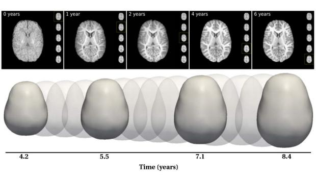
Better medical imaging, better data visualization, better healthcare

Longitudinal images of brains of children at risk or later diagnosed with autism spectrum disorder developed by the Visualization, Imaging, and Data Analytics (VIDA) Center at NYU
Researchers at the Visualization, Imaging, and Data Analytics (VIDA) Center at NYU work closely with domain experts in a variety of fields to apply the latest advances in computing to problems of critical societal importance, and some of their most exciting work is currently being conducted with medical personnel from NYU Langone.
Guido Gerig, an institute professor and chair of the Department of Computer Science and Engineering, is considered to be among the world’s foremost authorities in the field of medical image analysis, and at VIDA, he and his colleagues and students are tackling some of medicine’s most challenging puzzles.
A fellow of the American Institute for Medical and Biological Engineering (AIMBE), the Medical Image Computing and Computer-Assisted Intervention (MICCAI) Society, and the Institute of Electrical and Electronics Engineers (IEEE), he is making vast contributions to our understanding of autism, traumatic brain injury, glaucoma, drug exposure, and other conditions.
VIDA RESEARCH PROJECTS:
Novel Glaucoma Diagnostics for Structure and Function
Fellow researchers: Joel Schumann, chair of NYU Ophthalmology, director of NYU Langone Eye Center, and NYU Tandon professor of Biomedical Engineering and Electrical & Computer Engineering (principal investigator); Hiroshi Ishikawa, Professor at NYU Ophthalmology, Research Professor James Fishbaugh; Ph.D. candidate Sungmin Hong; Intern Guillaume Gisbert
Problem: Patients with glaucoma suffer damage to the optic nerve that can lead to loss of vision and even blindness; the disease is the leading cause of irreversible blindness in the world.
Research: Because glaucoma is most often associated with elevated intraocular pressure, researchers use optical coherence tomography (a technique pioneered by Schumann) to obtain images of the retina and eye nerve. This is followed by advanced image analysis to detect pathological changes in the optic nerve. Their overall goal is to find imaging biomarkers for very early changes in the optic nerve area, well before loss of vision can occur.
Hip Chondromics: Comprehensive Cartilage Characterization with MR Fingerprinting
Fellow researchers: Riccardo Lattanzi, associate professor of Electrical and Computer Engineering at Tandon and Radiology at Langone; Research Professor James Fishbaugh; Ph.D. candidate Batool Abbas
Problem: Femoroacetabular impingement (FAI) is a pathologic condition in which structural abnormalities of the femoral head-neck junction and/or acetabulum lead to a mechanical blockage in the hip joint that compromises range of motion. If the impingement is left untreated, it can cause cartilage damage and lead to hip osteoarthritis.
Research: Lattanzi, Gerig, and their colleagues aim to demonstrate the utility of multi-parametric quantitative magnetic resonance (MR) imaging to manage FAI and to develop the technology necessary to translate it into clinical routine.
Analysis of the Kinematic Motion of the Wrist from 4D MR Images
Fellow researchers: Associate Professor of Radiology Catherine Petchprapa, Lattanzi, Fishbaugh, and Abbas
Problem: The complex articulations of the wrist allow for a wide range of movement through the interactions of the eight carpal wrist bones, and while the study of this intricate system is vital to the understanding, diagnosis, and treatment of wrist abnormalities, static MRI and CT are both limited in their capacity and usability in the study of wrist kinematics of living human subjects.
Research: 4D MRI (dynamic MR imaging covering a motion sequence of wrist movement) provides an effective means of addressing those limitations, but it comes with its own set of challenges, such as low resolution. The NYU researchers are developing methods to effectively solve the challenges and to quantify the 3D kinematics of dynamic wrist data acquired from 4D MRI.
Investigation of Opioid Exposure and Neurodevelopment (iOPEN) project
Fellow researchers: Moriah Thomason of NYU Langone Child and Adolescence Psychiatry (principal investigator) and Fishbaugh
Problem: It is imperative to recruit and retain pregnant women and their children for long-term study of exposure to opioids in utero and how it affects subsequent neurodevelopment.
Research: The researchers are implementing and analyzing harmonized MRI acquisitions for placental, fetal, neonatal, and toddler MRI by means of a multi-site, standardized research protocol; their aim is to maximize data quality, usability, and integration across all sites to inform the design of Phase II studies.
While NYU Langone has provided a rich trove of fellow researchers, Gerig and others at VIDA also partner frequently with those from other academic institutions throughout the country, including Washington University, the University of North Carolina at Chapel Hill, the University of Pennsylvania, and the University of Alabama, as well as with corporate partners like software company Kitware, Inc.
Some of their collaborative projects involve studying, among other vital topics:
- very early brain development in infants with Down syndrome
- autism from infancy through school age
- the effects of cocaine and maternal behavior on brain development
- the quantitative analysis of microscopy tissue images of patients with age-related macular degeneration (AMD)

