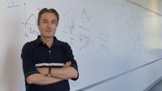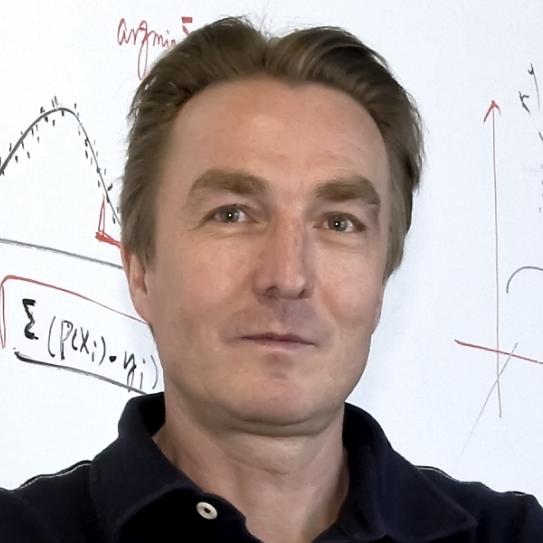
Guido Gerig is Department Chair and Institute Professor in the Department of Computer Science and Engineering and he is a professor of Biomedical Engineering. He also holds associated/affiliated appointments at NYU Courant CS, NYU Langone Child and Adolescent Psychiatry, Psychiatry and Radiology.
Guido Gerig has been named IEEE Fellow (class of 2019) and has also been appointed as a Fellow of the American Institute for Medical and Biological Engineering (AIMBE) in 2010.
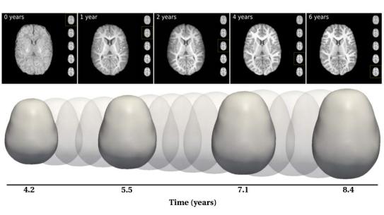
Starting image analysis with applications to satellite imaging, he became increasingly interested in driving problems from medicine, tackled in close multidisciplinary collaboration between medicine, engineering, and statistics. His research supports a number of clinical imaging research studies with novel, innovative image analysis methodologies related to segmentation, registration, atlas building, shape analysis, and image statistics. Driving clinical problems include research in autism, Down's syncdrome, eye diseases (Glaucoma, AMD), multiple sclerosis, Huntington's disease, studies of infants at risk for mental illness, and in general analysis of anatomical changes from normal due to disease, therapy and recovery. Gerig's research resulted in various new image analysis methodologies for nonlinear processing, multi-scale segmentation and shape analysis, some of them first and seminal to the field. Applications resulted in new clinical research discoveries such as vulnerability for schizophrenia, early diagnosis of autism based on differences in brain development trajectories, and correlation of shape atrophy with risk status in Huntington's. New tools and methods are developed as open source software and made available to the public, including teaching materials and hands-on training workshops.
Guido Gerig was previously USTAR Professor of Computer Science at the University of Utah (2007-2015) establishing the Utah Center for Neuroimage Analysis (UCNIA), Taylor Grandy Professor of Computer Science and Psychiatry at the University of North Carolina at Chapel Hill (1998-2007) launching the UNC Neuro Image Research and Analysis Laboratories (NIRAL), and Assistant Professor at ETH Zurich (1993-1998). Gerig holds several awards from Utah and UNC for Excellence in Teaching.
http://library.med.nyu.edu/api/publications?person=gg87
http://www.sci.utah.edu/scipubs/search.html?schTerm=G.+Gerig
Education
Swiss Federal Institute of Technology Zurich (ETH-Z) 1981
MSc,
Swiss Federal Institute of Technology Zurich (ETH-Z) 1987
PhD,
Experience
University of Utah
USTAR Professor
From: July 2007 to June 2015
University of North Carolina UNC
Taylor Grandy Professor
From: July 1998 to June 2007
Swiss Federal Institute of Technology ETH
Assistant Professor
From: July 1993 to June 1998
Publications
Journal and Conference Articles (peer reviewed)
NIH Pubmed/PMC list of publications:
http://www.ncbi.nlm.nih.gov/pubmed?term=Gerig%20G
NYU Health Science Library:
http://library.med.nyu.edu/api/publications?person=gg87
Affiliations
NYU Courant Institute, Computer Science
NYU Radiology, Center for Biomedical Imaging
NYU Child and Adolescence Psychiatry
Awards
- 2019: IEEE Fellow (class of 2019)
- 2010: Elected Fellow of the American Institute for Medical and Biological Engineering (AIMBE)
- 2010 Dean's letter University of Utah for Excellence in Teaching
- 2009: Fellow of the Medical Image Computing and Computer-Assisted Intervention (MICCAI) Society
- 2009: ISMRM (Int Society for Magnetic Resonance in Medicine): Outstanding Teacher Award
- 2006, 2004, 1999: Student Teaching Awards, UNC Chapel Hill, Dept. Computer Science
Information for Mentees
About me: I joined Tandon 2015 after 5 years at ETH Zurich, 8 years at UNC-Chapel Hill and 8 years at U of Utah / was CSE chair 2016 - 2020 / joint appointment CSE/BME
Research News
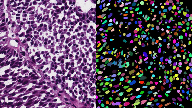
Microscopy image segmentation via point and shape regularized data synthesis
In contemporary deep learning-based methods for segmenting microscopic images, there's a heavy reliance on extensive training data that requires detailed annotations. This process is both expensive and labor-intensive. An alternative approach involves using simpler annotations, such as marking the center points of objects. While not as detailed, these point annotations still provide valuable information for image analysis.
In this study, researchers from NYU Tandon and University Hospital Bonn in Germany assume that only point annotations are available for training and present a novel method for segmenting microscopic images using artificially generated training data. Their framework consists of three main stages:
1. Pseudo Dense Mask Generation: This step takes the point annotations and creates synthetic, detailed masks that are constrained by shape information.
2. Realistic Image Generation: An advanced generative model, trained in a unique way, transforms these synthetic masks into highly realistic microscopic images while maintaining consistency in object appearance.
3. Specialized Model Training: The synthetic masks and generated images are combined to create a dataset used to train a specialized model for image segmentation.
The research was led by Guido Gerig, Institute Professor of Computer Science and Engineering and Biomedical Engineering, alongside PhD students Shijie Li and Mengwei Ren, as well as Thomas Ach at University Hospital Bonn. The three NYU Tandon researchers are also members of the Visualization and Data Analytics (VIDA) Research Center.
The researchers tested their method on a publicly available dataset and found that their approach produced more diverse and realistic images compared to conventional methods, all while maintaining a strong connection between the input annotations and the generated images. Importantly, when compared to models trained using other methods, their models, trained on synthetic data, outperformed them significantly. Moreover, their framework achieved results on par with models trained using labor-intensive, highly detailed annotations.
This research highlights the potential of using simplified annotations and synthetic data to streamline the process of segmenting microscopic images, potentially reducing the need for extensive manual annotation efforts. The research, in collaboration with the Ophthalmology department at University Hospital Bonn, is a first step in a collaboration to finally process three dimensional retinal cell images of the human eye from subjects diagnosed for age-related macular degeneration (AMD), a leading cause of vision loss in older adults.
The code for this method is publicly available for further exploration and implementation.
“Microscopy Image Segmentation via Point and Shape Regularized Data Synthesis.” S Li, M Ren, T Ach, G Gerig. arXiv preprint, arXiv:2308.09835, 2023.


